Invented by Pezzotti; Nicola, Wenzel; Fabian, Van den Brink; Johan Samuel, Weese; Rolf Jürgen, Flaeschner; Nick, Doneva; Mariya Invanova, Ewald; Arne
Welcome! Today, we will explore a new patent application for a medical system that aims to make MRI follow-up imaging more affordable and accessible. We will break down what the invention is, why it matters, and how it works. This article will help you understand the context, the science behind the idea, and the key parts of the invention. Let’s get started.
Background and Market Context
MRI, or Magnetic Resonance Imaging, is a tool that helps doctors see inside the body without surgery. It uses powerful magnets and radio waves to make detailed pictures. These pictures help doctors find problems in the brain, spine, joints, organs, and more. Because MRI is safe and does not use harmful radiation, it is one of the best ways to check for diseases, injuries, or tumors.
Even though MRI is very useful, it is also expensive. The main reason is the strong magnets inside the machines. Hospitals spend a lot of money to buy, install, and keep these machines working. The stronger the magnet, the clearer the images, but the higher the cost. This means that many smaller clinics and hospitals, especially in rural or less wealthy areas, cannot afford high-strength MRI machines. Patients in these places might have to travel far for scans, which is not always possible.
There is a way to make MRI cheaper: use magnets that are not as strong. These are called low-field MRI machines. They cost less to make and keep running. But there is a problem—images from these machines are not as sharp. The details are blurrier, and it is harder to spot small changes or problems. Doctors worry that if images are not clear, they might miss something important.
But what if we did not need high-detail images every time? When a patient first gets sick, or when doctors are trying to make a diagnosis, they need the best images. But for follow-up scans—when checking if something has changed, like if a tumor has grown or shrunk—maybe less detail is okay, as long as the changes can still be tracked. This is where the new invention comes in.
The patent application describes a system that lets clinics use low-field MRI machines for follow-up scans. The idea is simple but powerful: use a high-detail scan from a big hospital as a “baseline,” and then use cheaper, low-detail scans for later checkups. The system compares the new images to the old ones, looking for big changes. If something is different by a certain amount, it sends a warning to the doctor or patient. This could make follow-up care easier, faster, and more affordable for many people.
This solution could open the door for more widespread MRI use, especially in places where cost is a big barrier. It could also save time for patients, reduce the burden on busy hospitals, and help doctors catch problems early without waiting for high-end machines to be available.
Scientific Rationale and Prior Art
Let’s talk about the science behind MRI and why low-field images are usually not good enough. MRI works by using a strong magnetic field to align tiny particles in your body, called nuclei. When the machine sends a radio wave, these particles “wiggle” and send back signals. The machine listens to these signals and turns them into images. The stronger the magnetic field, the more the particles line up, and the clearer the signals. This means better, sharper pictures.
High-field MRI machines (those with fields above 1 Tesla) give the best images. They can show tiny differences between healthy and sick tissue. Low-field machines (with fields less than 0.6 Tesla, and sometimes as low as 0.2 Tesla) are cheaper but the images are fuzzier. The signals from the body are weaker, and there is more “noise” (random stuff that makes the image messy). It is harder to see small tumors, thin tissues, or small changes over time.
In the past, some researchers have tried to make low-field MRI more useful. For example, a 2016 journal article by Wu, Chen, and Nayak talked about ways to simulate what low-field images would look like, using data from high-field scans. This helped scientists understand what they were missing, but it did not solve the problem for real patients.
Another patent (US2018/0025466) described ways to make sure follow-up MRI scans were taken in the same spot and at the same angle as the first scan. This is important because if you are not looking at the same place, you cannot compare the images. But this patent did not focus on using low-field machines for follow-ups or on how to compare images of different quality.
The challenge is that low-field images look different from high-field ones, not only because of lower sharpness but also because of changes in how tissues appear. The way the body’s water and fat respond to the magnets is not exactly the same. This means that just looking at a low-field image and a high-field image side by side may not be enough. Doctors could miss a growing tumor or think something has changed when it really hasn’t.

The new patent addresses this problem by using computer systems and smart software to “bridge the gap.” It uses the baseline measurements from a high-field scan to guide what to look for in the low-field scan. The system knows where to look (using landmarks in the body), what size or shape to expect, and even how much change is too much. It can use advanced tools like lookup tables, expert systems, or even trained neural networks (a type of artificial intelligence) to help make sense of the fuzzier images. The computer does the hard work of measuring and comparing, and only alerts the doctor if something important has changed.
This approach builds on the work done before but goes further. It does not just try to make low-field images look better. Instead, it builds a smart system that uses all the information from the first scan, the machine settings, and the patient’s own anatomy, to get the most out of cheaper follow-up scans. It is a practical answer to a real-world problem.
Invention Description and Key Innovations
Now let’s break down what the invention does and what makes it new and special.
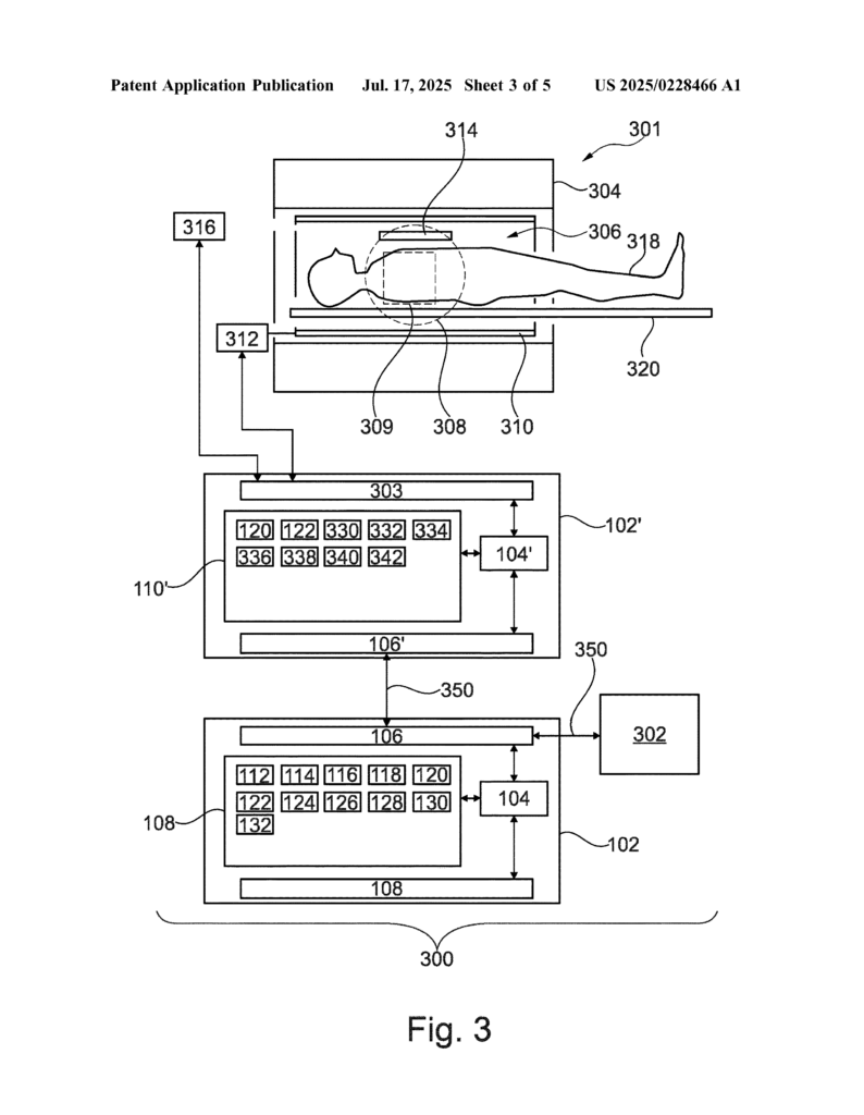
At its heart, the invention is a medical system made up of a few main parts:
– A computer system with memory, network connections, and smart software (machine instructions).
– A low-field MRI machine that can take basic scans.
– (Sometimes) a high-field MRI machine for the first “baseline” scan.
Here is how the system works, step by step:
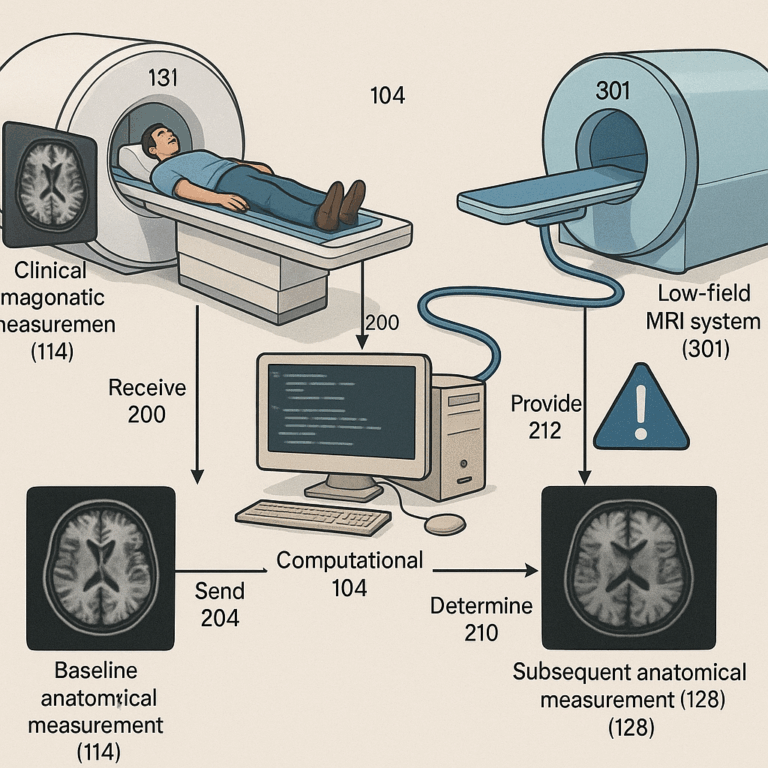
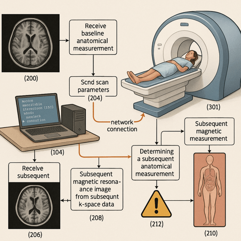
First, the system gets a baseline anatomical measurement. This means that after a patient’s first high-quality MRI scan (at a hospital or imaging center), the system stores important details: maybe the size of a tumor, the width of an organ, or the distance between two landmarks in the body. It also keeps track of extra information, called scan metadata. This includes where the scan was taken on the body, the settings used, and the strength of the magnet.
When it is time for a follow-up scan, the system uses all this saved information to plan the new scan. It sends instructions (called scan parameters) over a network to a low-field MRI machine. These instructions tell the machine exactly what to do—where to scan, how sharp the image needs to be, and what to focus on. The instructions might be chosen from a list, picked by an expert system, or even decided by a neural network that has learned from past scans.
The low-field MRI machine takes the new scan and sends the raw data (called k-space data) back to the computer system. The computer then rebuilds this data into a new MRI image.
Now comes the smart part. The computer uses a segmentation module—a special piece of software that finds the right spot in the new image and measures the same thing as before (like the size of a tumor). This module could be a simple algorithm, a set of rules, or a neural network trained to work with blurry images and still get the right answer.
Once the measurement is done, the computer compares it to the baseline. If the difference is bigger than a certain amount (a threshold set by doctors), the system sends an alert. This warning could go to a doctor, a nurse, or even the patient. It might come as an email, a message on a phone, or a note in the patient’s record. The system could also tell the MRI machine to try again if something seems off.
There are a few more clever features:
– The system can work as a cloud service, helping many clinics and MRI machines at once.
– The segmentation and measurement can adjust to different kinds of images and scan settings.
– As the system is used more, it gets smarter—by storing data from each scan, it can improve its settings for next time, either through tables or by training its neural networks.
– The warning system is flexible. It can send messages to people or to machines, or even suggest new scans or health actions for the patient.
The invention also describes ways for the MRI machine itself to help. For example, before the real scan, the machine might take a quick “survey” scan to find the right spot. It can use this survey to make sure the follow-up scan matches the first one, even if the patient is lying a little differently. This makes sure the images can really be compared.
The key innovations here are:
– Using low-field MRI machines for follow-up scans, guided by a high-quality baseline.
– Smart software that takes care of matching, measuring, and comparing, even when the new images are less clear.
– A flexible system that can be used in many places, helping make MRI follow-up cheaper and more available.
– Learning from each scan to get better over time, using AI and data storage.
All of these steps help solve the big problem: how to keep track of changes in the body, without needing expensive, high-end MRI machines every time. By focusing on what matters (the measurement, not the image itself), and letting computers handle the tricky comparison, the invention brings MRI follow-up to more places and more people.
Conclusion
This patent application offers a new way to make MRI follow-up imaging less expensive and more accessible. By using high-quality baseline scans from standard MRI and then switching to affordable low-field MRI for future check-ups, the system helps clinics save money while still keeping patients safe. The real magic comes from the smart computer software that knows how to measure, compare, and alert when something important has changed—even when the new images are not as clear.
This approach could help small clinics, rural hospitals, and busy health systems provide better care for more people, without waiting for costly equipment. By making follow-up imaging as easy as possible, this invention can help catch health problems early, improve patient outcomes, and bring advanced medical care to new places. As the technology keeps learning and growing, it could become a key part of how doctors track diseases, monitor recovery, and keep patients healthy around the world.
Click here https://ppubs.uspto.gov/pubwebapp/ and search 20250228466.